Abstract
May-Thurner Syndrome (MTS) also known as iliac vein compression syndrome, is a congenital anatomical variant, that results from the extrinsic compression of the left common iliac vein by the right iliac artery with resultant formation of left venous thrombosis. The compression may cause leg swelling, varicosities, deep vein thrombosis (DVT), chronic venous stasis ulcers or more serious complications such as pulmonary embolism. MTS usually asymptomatic until DVT occurs. Here we report an atypical case where MTS was resulted from compression of left common iliac vein (LCIV) by the left common iliac artery (LCIA).
Keywords
May-Thurner Syndrome (MTS) deep vein thrombosis (DVT) Cockett syndrome iliocaval compression syndrome unilateral leg swelling
Introduction
May-Thurner syndrome, also known as iliac vein compression syndrome, Cockett syndrome, or iliocaval compression syndrome is caused when the left iliac vein is compressed by the right iliac artery, which increased the risk of deep vein thrombosis (DVT) in the left leg (1-3). A history of persistent left lower extremity swelling with or without deep venous thrombosis in a woman between the 2nd and 4th decades of life, without an obvious cause, is highly suggestive of May--Thurner syndrome. The clinical suspicion can be confirmed with CT and iliac venography (4).
May-Thurner syndrome (MTS) was first described by Rudolf Virchow in the early 1850s when he observed an increased incidence of the left iliofemoral vein compression by the right common iliac artery in a cadaveric study of patients with left iliofemoral thrombosis (5). This condition was later described by May and Thurner in 1957 as autopsies on cadavers showed this same anatomic variant with intraluminal fibrous band in the left common iliac vein. The condition later became widely known by their names.
The estimated prevalence of MTS in the general population is 14%-32%. It is however implicated in only 2%-5% of cases of lower limb deep vein thrombosis (6,7). Risk factors for MTS include female sex, multiparity, scoliosis, oral contraceptive use, cumulative radiation and hypercoagulable disorders (8,9,10). Development of this syndrome occurs in 3 stages; asymptomatic left common iliac vein compression, formation of a venous spur and finally left lower extremity DVT. Most cases patients with MTS never develop DVT (11). Very few existing literatures have described recurrent DVT in the setting of May-Thurner in young males.
Case report
A 50-year-old female patient presented at internal medicine department with the complain of 4 days history of left lower limb swelling and tenderness around thigh area. No pain around left knee joint area. No fever, no chest pain or shortness of breath. On examination significant lower limb oedema was found (Right leg 39 cm and left leg 45 cm). Clinical diagnosis was left lower limb DVT provoked, post left knee diagnosed arthroscopy two weeks back.
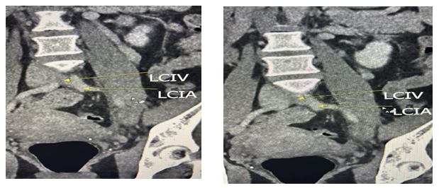
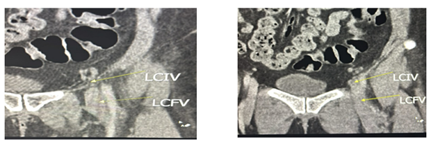
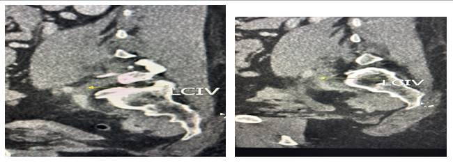
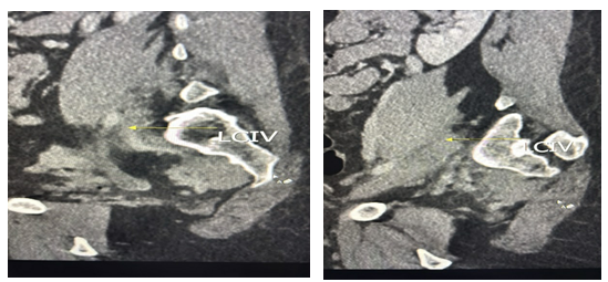
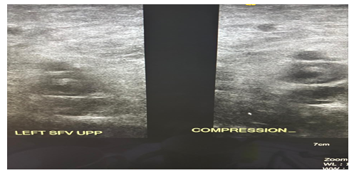
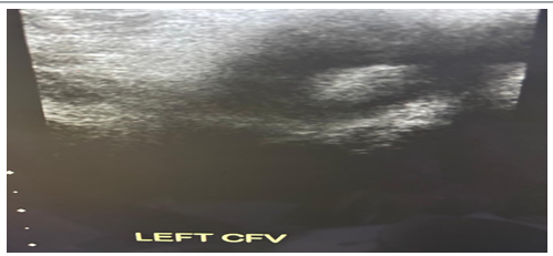
U/S venous Doppler was done and findings were extensive DVD in the left lower limb from the common femoral vein all the way down to the popliteal vein. CT pulmonary angiography showed no evidence of PE. CT TAP showed that the left common iliac artery compressed the left common iliac vein against the lumbar spine causing the deep vein thrombosis (DVT) (Fig1 – 4). U/S scan also confirmed thrombus in left superficial femoral vein (LSFV) and left common femoral vein (LCFV) (Fig. 5-6) The case was diagnosed as a variant of May Thurner Syndrome.
Treatment was given for long term anticoagulant and referred to vascular surgeon for consideration of endovascular stenting.
Discussion
In young patient presenting with DVT, a detailed history, physical examination, and diagnostic are warranted. May-Thurner syndrome is caused by compression of the left common iliac vein by the right common iliac artery. However, in some atypical cases the compression may cause by left common iliac artery also as demonstrated in this case. MTS is not an infrequent source of venous abnormalities in the left lower extremity.
The overlying artery appears to induce a partial obstruction of the vein in two ways: By its anatomic orientation with subsequent physical entrapment of the left common iliac vein and by extensive intimal hypertrophy of the vein resulting from the chronic pulsatile force of the right common iliac artery. This condition has been estimated to occur in 2-5% of patients who undergo evaluation for lower extremity venous disorders.
Reported findings show it is three to eight times more frequent in the left side than on the right. Iliac vein compression should be diagnosed prior to the onset of iliofemoral venous thrombosis and venous insufficiency syndrome. An iliac venogram obtained via femoral artery is the diagnostic test of choice because it can be demonstrating the compression itself and because pressure gradient measurement can be performed to confirm the hemodynamic significance. Contrast CT abdomen at distal abdominal aortic level can also demonstrate the extrinsic compression caused by the right common iliac artery.
MTS is a progressive disease with long term disabling complications. This type of obstruction may cause leg swelling, varicosities, DVT, chronic venous stasis ulcers, or more serious complications, such as pulmonary embolism or phlegmasia cerulea dolens.
MTS requires intervention when symptomatic. The mainstay of management of MTS is the removal of the clot with pharmaco-chemical thrombolysis and mechanical thrombectomy to prevent post thrombotic syndrome. Catheter directed thrombolysis with anticoagulation in iliofemoral DVT is superior to anticoagulation alone. Anticoagulation on its own is insufficient to manage MTS with DVT. Multiple surgical treatment options have been advocated such as vein patch angioplasty with excision of the intraluminal bands, division of the right common iliac artery and relocation behind the left common iliac vein or inferior vena cava. Contralateral saphenous vein graft bypass to the ipsilateral common femoral vein with creation of a temporary arteriovenous fistula. In recent years endovascular stent placement has been reported.
The diagnosis of MTS is made by imaging modalities include Doppler USG, Plethysmography, CT venography, MR venography and conventional venography. Contrast venography is considered the gold standard modality for MTS (11). In our study, however, the imaging modalities used were USG doppler and CT angiography. USG is often the first-line technique used in diagnosing DVT. Although it is the most common imaging modality. It has several limitations in the diagnosing MTS. These limitations include technical difficulties in assessing the iliac veins and inferior vena cava and false positive high flow velocity in the common iliac vein which may indicate obstruction or compression (5,12). In our case, USG demonstrated DVT but failed to identify the compression of the iliac veins.
CT venography has a high sensitivity and specificity to detect MTS. It has other advantages including its ability to rule out extrinsic compressions like lymphadenopathy and hematoma, identify acute DVT, and outline collateral pathways. CT also eliminates the need for technical expertise. It has limitations in pregnancy due to radiation dose and can overestimate the degree of compression in dehydration patients (6,13,14). In our patient CT with contrast accurately diagnosed MTS hence the use of this imaging modality in this condition is justified.
Conclusion
MTS results in LCIV compression, increasing the risk of DVT and PE. MTS in young, healthy women with unexplained left sided DVT, emphasizing the importance of early diagnosis for effective management and prevention of further issues. Unlike the typical presentation of MTS involving older patients with clotting disorders, this case provides a unique presentation due to the patient’s young age and extensive DVT. MTS should be taken into consideration if there is persistent edema of the left leg, especially in young women. The mechanical compression should be recognized prior to the onset of deep venous thrombosis and venous insufficiency. Endovascular treatment with stent placement is an alternative to direct surgical repair that yields excellent short-term results.
Conflict of interest: None.
References
- May R, Thurner J. The cause of the predominantly sinistral occurrence of thrombosis DOI ↗ Google Scholar ↗
- Cockett FB, Thomas ML. The iliac compression syndrome. Br J Surg. 1965;52:816–21. doi: . [DOI] [PubMed] [Google Scholar] DOI ↗ Google Scholar ↗
- Taheri SA, Williams J, Powell S, et al. Iliocaval compression syndrome. Am J Surg. 1987;154:169–72. DOI ↗ Google Scholar ↗
- Cil BE, Akpinar E, Karcaaltincaba M, Akinci D. Case 76: May-Thurner syndrome. Radiology. 2004;233:361–5. DOI ↗ Google Scholar ↗
- Harbin M.M., Lutsey P.L. May-Thurner syndrome: history of understanding and need for defining population prevalence. J Thromb Haemost. 2020;18(3):534–542. DOI ↗ Google Scholar ↗
- Mangla A., Hamad H. StatPearls. StatPearls Publishing; Treasure Island (FL): 2022. May-Thurner Syndrome. DOI ↗ Google Scholar ↗
- Kalu S., Shah P., Natarajan A., Nwankwo N., Mustafa U., Hussain N. May-Thurner syndrome: a case report and review of the literature. Case Rep Vasc Med. 2013;2013 DOI ↗ Google Scholar ↗
- Marston W., Fish D., Unger J., Keagy B. Incidence of and risk factors for iliocaval venous obstruction in patients with active or healed venous leg ulcers. J Vasc Surg. 2011;53(5):1303–1308. DOI ↗ Google Scholar ↗
- Oteros Fernández R., Bravo Rodríguez F., Delgado Acosta F., González Barrios I. Síndrome de May- Thurner y cirugía de escoliosis [May-Thurner syndrome and surgery for scoliosis] Radiologia. 2008;50(3):245–247. DOI ↗ Google Scholar ↗
- Zander K.D., Staat B., Galan H. May-Thurner Syndrome resulting in acute iliofemoral deep vein thrombosis in the postpartum period. Obstet Gynecol. 2008;111(2 Pt 2):565–569. DOI ↗ Google Scholar ↗
- Brinegar K.N., Sheth R.A., Khademhosseini A., Bautista J., Oklu R. Iliac vein compression syndrome: clinical, imaging, and pathologic findings. World J Radiol. 2015;7(11):375–381. DOI ↗ Google Scholar ↗
- Metzger P.B., Rossi F.H., Kambara A.M., Izukawa N.M., Saleh M.H., Pinto I.M., et al. Criteria for detecting significant chronic iliac venous obstructions with duplex ultrasound. J Vasc Surg. Venous Lymphat Disord. 2016;4(1):18–27. DOI ↗ Google Scholar ↗
- Wu W.L., Tzeng W.S., Wu R.H., Tsai W.L., Chen M.C., Lin P.C., et al. Comprehensive MDCT evaluation of patients with suspected May-Thurner syndrome. AJR Am J Roentgenol. 2012;199(5):W638–W645. DOI ↗ Google Scholar ↗
- Ibrahim W., Al Safran Z., Hasan H., Zeid W.A. Endovascular management of May-Thurner syndrome. Ann Vasc Dis. 2012;5(2):217–221. DOI ↗ Google Scholar ↗