Abstract
Abstract Background: Malaria and dengue fever are both endemic in Kenya and share overlapping clinical features, which can delay recognition of coinfection. Reports of concurrent Plasmodium and Dengue virus infection have increased across Sub-Saharan Africa in the last decade. Objective: We highlight a case of a Kenyan soldier who developed severe, multi-organ malaria-dengue coinfection after returning from the coast to Wajir (Northeastern Kenya), and discuss the epidemiological, diagnostic, and management considerations it raised. Case presentation: We describe a 32-year-old male soldier who developed fever, persistent vomiting, retro-orbital headache, myalgia, and low back pain five days after returning to Wajir following a three-week deployment to the Kenyan coast. Investigations confirmed concurrent falciparum malaria and dengue fever complicated by severe thrombocytopenia (platelet count 36 × 10⁹/L), acute kidney injury (creatinine 137.04 µmol/L), and transaminitis. He was managed with intravenous artesunate, intravenous fluids, empirical antibiotics, antiemetics, and close monitoring for bleeding tendencies. Renal function normalized within 24 hours, and thrombocytopenia and hepatic derangement resolved progressively, allowing transition to oral artemether-lumefantrine by day 2 of admission and discharged. Conclusion: This case illustrates that malaria-dengue coinfection can present with multi-organ derangement even outside classically high-transmission zones, underscoring the value of dual rapid diagnostic testing and early WHO-guideline-directed severe malaria treatment, in febrile patients moving between ecologically distinct, co-endemic regions such as coastal and northern Kenya.
Keywords
Keywords: Malaria Dengue fever Coinfection Acute kidney injury Thrombocytopenia.
Introduction
Malaria and dengue fever remain the two most important causes of acute undifferentiated febrile illness in the tropics and subtropics. Both are transmitted by mosquitoes, circulate widely across sub-Saharan Africa, and can present with , making them difficult to distinguish on clinical grounds alone. Historically, dengue and malaria coinfection was considered rare, but a growing number of cross-sectional studies and case reports from Africa, Asia and Latin America indicate that the two infections coexist more often than previously appreciated, particularly in regions where their respective mosquito vectors overlap.
A recent meta-analysis estimated a pooled prevalence of malaria and acute dengue virus coinfection among febrile patients in Africa of 4.2%, with East Africa contributing a substantial share of reported cases and with prevalence rising steadily over the past decade as dengue surveillance capacity has improved (1). A separate systematic review found that roughly one-third of patients with confirmed Plasmodium-dengue coinfection went on to develop features of severe malaria, and that coinfected patients carried a significantly higher risk of severe disease than those with dengue monoinfection, with bleeding, jaundice and hypotension the most frequently reported severe complications (2).
alaria transmission the Lake Victoria basin and along the coastal strip, the arid and semi-arid counties of northern Kenya, including Waji(3). engue, has been recognized on the Kenyan coast since an outbreak in Mombasa in 1982, with subsequent large outbreaks confirmed in 2011, 2013–2014 and 2017(4). ncreasing climatic variability and population mobility have since been associated with rising malaria risk even in traditionally low-transmission northern counties, blurring the geographic boundaries that once separated these two vector-borne diseases (3).
Military and other security who are redeployed between ecologically distinct regions within short intervals represent a population particularly susceptible to importing infection from one endemic zone into another, and to acquiring coinfection while still in the incubation period of a first illness. We report the case of a Kenyan soldier who developed severe, multi-organ malaria-dengue coinfection after returning from the coast to Wajir, and discuss the epidemiological, diagnostic and management considerations it raise
Case History
32-year-old male soldier, presented with a five-day history of fever, nausea severe vomitingdiffuse joint pains, chills and generalized body malaise. He described intermittent headaches with severe retro-orbital pain, mild intermittent right and left upper-quadrant abdominal pain. convulsions, bleeding tendencies, cough, chest pain or .
been deployed to the Kenyan coast weeks before symptom onset
Physical Examination
On examination, with a temperature of 38.0°C without conjunctival pallor. lood pressure 113/68 mmHg, pulse rate 98 beats/min, oxygen saturation 98% on room air, and random blood sugar 4.8 mmol/L. Abdominal examination revealed examination
Investigations
Baseline , biochemical and rapid diagnostic testing on admission
Diagnosis
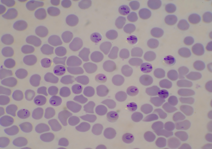
of severe falciparum malaria in coinfection with dengue fever was established, complicated by severe thrombocytopenia, acute kidney injury and hepatic . in keeping with World Health Organization severity criteria for falciparum malaria (5). The patient was admitted for inpatient management.
Management and Clinical Course
ntravenous artesunate was commenced at a dose of 2.4 mg/kg, administered at 0, 12 and 24 hours. Supportive management included intravenous fluid resuscitation with Ringer's lactate alternating with 5% dextrose, intravenous paracetamol for antipyresis and analgesia, and intravenous ondansetron for refractory vomiting. Empirical intravenous ceftriaxone 2 g once daily was started for five days. Given the severe thrombocytopenia, the patient was monitored four-hourly for vital signs, random blood sugar, mental status, and any clinical evidence of bleeding.
reported significant clinical improvementThere was no evidence of bleeding. epeat investigations showed (platelet count 42 × 10⁹/L), but renal function and electrolytes had normalized anagement was continued unchanged.
| Parameter | Admission (Day 0) | Day 1 | Day 2 | Day 3 |
| Platelet count (×10⁹/L) | 36 | 42 | 86 | 106 |
| (g/dL) | 15.9 | 14.9 | 13.9 | 13.4 |
| 137.04 | 123 | 85 | normal | |
| Electrolytes | Na⁺ 132.53, K⁺ 3.32 | Na⁺ 138, K⁺ 3.92 | Na⁺ 140, K⁺ 4.1 | normal |
| Clinical status | Sic, febrile, dehydrated | Improved, afebrile, mild scleral jaundice | Marked improvement | Clinically normal |
Discussion
Epidemiological Context
This case is notable for the geographic trajectory of the patient's exposure. Coastal Kenya has long been recognized as an area of continuous malaria transmission and has experienced recurrent, well-documented dengue outbreaks since 1982, with entomological surveys confirming established Aedes aegypti populations driving Mombasa outbreaks in 2013–2014 (4). Wajir and other arid northern counties, by contrast, have traditionally been classified as low or seasonal transmission areas for malaria and have historically featured in dengue surveillance, although rising temperatures, altered rainfall patterns and population movement have been associated with expanding malaria risk in these areas in recent years (3). The three-week interval between the patient's coastal deployment and symptom onset, together with the incubation periods typical of falciparum malaria (roughly 7–30 days) and dengue virus (typically 4–14 days), with acquisition of both infections while on the coast, with clinical manifestation only after his arrival in Wajirillustrat how troop and movement can effectively relocate coinfection risk
Clinical and Diagnostic Overlap
Malaria and dengue share a broad range of early symptoms fever, chills, headache, myalgia, arthralgia and vomiting differentiation unreliable. This overlap underdiagnosis of coinfection in malaria-endemic African settings, where a positive malaria test is often accepted as sufficient explanation for febrile illness without further evaluation for dengue or other arboviral disease (1). Retro-orbital pain, as seen in this patient, is a classically dengue-associated symptom, while lower back and joint pain, chills and hepatosplenomegaly can be seen in either infection, further blurring the clinical picture. This reinforces the recommendation, across the coinfection literature, that clinicians in co-endemic or newly co-endemic areas should pursue dual testing in patients with atypically severe or prolonged febrile illness, rather than assuming a single diagnosis (1, 7).
Severity and Pathophysiological Considerations
The combination of severe thrombocytopenia, acute kidney injury and hepatic dysfunction observed in this patient is consistent with patterns described in other reports of malaria-dengue coinfection. A systematic review and meta-analysis found that approximately one-third of coinfected patients developed features of severe malaria, with jaundice and bleeding among the most common severe complications, and that coinfection carried a significantly higher risk of severe disease than dengue monoinfection alone (2). matched-pair French Guiana found that patients with dengue-malaria coinfection had significantly deeper thrombocytopenia and lower suggest a synergistic effect on platelet consumption, possibly reflecting combined dengue-associated marrow platelet destruction malaria-associated splenic sequestration (8).
The clinical trajectory in this case was considerably more several published reports of severe coinfection. fatal case of cerebral malaria with dengue coinfection in China progressed to multi-organ dysfunction and septic shock despite antimalarial treatment (9), while a case from Nepal describing dengue with subsequently diagnosed Plasmodium vivax coinfection showed a more protracted, course marked by persistent fever, hyperbilirubinemia and renal impairment (10). Similarly, a Malaysian report of concurrent Plasmodium knowlesi and dengue infection highlighted the risk of a missed or delayed second diagnosis when a first pathogen is identified early (11). By comparison, this patient's renal function normalized within 24 hours and his platelet count and hepatic markers improved steadily from day 1, an outcome most plausibly attributable to presentation and treatment initiation within the first week of illness, before more advanced organ dysfunction could develop.
Management Rationale
Intravenous artesunate at 2.4 mg/kg administered at 0, 12 and 24 hours remains the WHO- and CDC-recommended first-line treatment for severe falciparum malaria in adults, regardless of the infecting Plasmodium species, and is associated with substantially lower mortality than parenteral quinine (5, 6). Transition to a full oral artemisinin-based combination course, once the patient has completed at least 24 hours of parenteral treatment and can tolerate oral intake, is standard practice; current guidance also supports an extended, five-day course of artemether-lumefantrine as follow-on therapy for falciparum malaria(6). Fluid management with alternating crystalloid and dextrose infusions, together with close monitoring of blood glucose, addressed the risks of dehydration and artesunate- or malaria-associated , while empirical ceftriaxone was reasonable pending exclusion of an alternative or co-existing bacterial cause of febrile illness in a region where enteric fever and other bacterial infections remain important differentials, although antimicrobial stewardship considerations would support early discontinuation once bacterial infection is excluded onitoring was appropriate given the patient's severe thrombocytopenia and the recognized risk of complications in dengue-malaria coinfection (2).
Limitations
This case relied on rapid diagnostic testing for both malaria and dengue confirmatory microscopy with parasite density quantification, dengue NS1/IgM serology or molecular testing, and dengue serotype could not be determined he findings cannot be generalized taken to minimize the potential severity of this coinfection, which has been associated with fatal elsewhere in the literature (9).
Conclusion
This case demonstrates severe, multi-organ malaria-dengue coinfection even a patient outside a classically high-transmission zoneIt reinforces the need for clinicians managing febrile illness in mobile populations including military and frequent to maintain a high index of suspicion for coinfection, pursue dual rapid diagnostic testing for malaria and dengue where plausible, and apply WHO-guideline-directed severe malaria treatment promptly. Early recognition and treatmepoorer outcomes reported in more advanced or delayed presentations.
Declarations
Consent for publication:nformed consent was obtained from the patient for publication of this case and any accompanying clinical details.
Conflict of interest: The authors declare no conflict of interest.
Funding: No funding was received for this case report.
References
- Gebremariam TT, Schallig HDFH, Kurmane ZM, Danquah JB. Increasing prevalence of malaria and acute dengue virus coinfection in Africa: a meta-analysis and meta-regression of cross-sectional studies. Malar J. 2023;22:300. DOI ↗ Google Scholar ↗
- Kotepui M, Kotepui KU, Milanez GDJ, Masangkay FR. Prevalence of and risk factors for severe malaria caused by Plasmodium and dengue virus co-infection: a systematic review and meta-analysis. Infect Dis Poverty. 2020;9:134. DOI ↗ Google Scholar ↗
- Omondi CJ, Onguru D, Kamau L, Nanyingi M, Ong'amo G, Estambale B. Perennial transmission of malaria in the low altitude areas of Baringo County, Kenya. Malar J. 2017;16:257. Google Scholar ↗
- Lutomiah J, Barrera R, Makio A, Mutisya J, Koka H, Owaka S, Koskei E, Nyunja A, Eyase F, Coldren R, Sang R. Dengue outbreak in Mombasa City, Kenya, 2013–2014: Entomologic investigations. PLoS Negl Trop Dis. 2016;10(10):e0004981. DOI ↗ Google Scholar ↗
- World Health Organization. Guidelines for the Treatment of Malaria. 3rd ed. Geneva: WHO; and Severe Malaria Observatory / Medicines for Malaria Venture. Injectable Artesunate dosing guidance. Available from: https://severemalaria.org/node/100 Google Scholar ↗
- Centers for Disease Control and Prevention. Treatment of Severe Malaria: Clinical Guidance for Health Care Providers. Atlanta: CDC. Available from: https://www.cdc.gov/malaria/hcp/clinical-guidance/treatment-of-severe-malaria.html Google Scholar ↗
- Alsedig K, Eldigail MH, Elduma AH, Elaagip A, Altahir O, Siam HA, et al. Prevalence of malaria and dengue co-infections among febrile patients during dengue transmission season in Kassala, eastern Sudan. PLoS Negl Trop Dis. 2023;17(10):e0011660. Google Scholar ↗
- Epelboin L, Hanf M, Dussart P, Ouar-Epelboin S, Djossou F, Nacher M, Carme B. Is dengue and malaria co-infection more severe than single infections? A retrospective matched-pair study in French Guiana. Malar J. 2012;11:142. Google Scholar ↗
- Zhao Y, Wu X, Liao F. Severe Cerebral Falciparum Malaria with Dengue Coinfection: A Case Report. Iran J Parasitol. 2018;13(2):323–327. Google Scholar ↗
- Gautam A, Aryal U, Bhandari S, Pradhan S, Bhattarai U, Mishra A, Sharma SK. Dengue and malaria coinfection: the first case report in Nepal. Oxf Med Case Reports. 2022;2022(3):omac022. Google Scholar ↗
- Chong SE, Mohamad Zaini RH, Suraiya S, Lee KT, Lim JA. The dangers of accepting a single diagnosis: case report of concurrent Plasmodium knowlesi malaria and dengue infection. Malar J. 2017;16:2. Google Scholar ↗